What is Osteoid Osteoma?
Osteoid Osteoma is a benign tumour of the bone occurring usually in children and young adults, though it can affect any age group. It does not spread to other parts of the body, though the pain & discomfort caused by it can be too much.
Is osteoid osteoma Cancerous?
No, Osteoid osteoma is not cancerous and hence doesn’t cause any threat to the life of the patient.

Is there any gender predilection for osteoid osteoma?
Osteoid osteoma tends to affect males up to three times more than females.
What are the common symptoms of Osteoid Osteoma?
Pain is the most common and disabling symptom of Osteoid Osteoma. Pain is usually severe and dull aching. Typically, pain is more in the night severe enough to wake up the patient from sleep. Usually this pain lingers on for months to years before patient seeks expert advise, since the pain goes away with over the counter pain killers.
Osteoid osteoma close to growth plates can lead to skeletal asymmetry
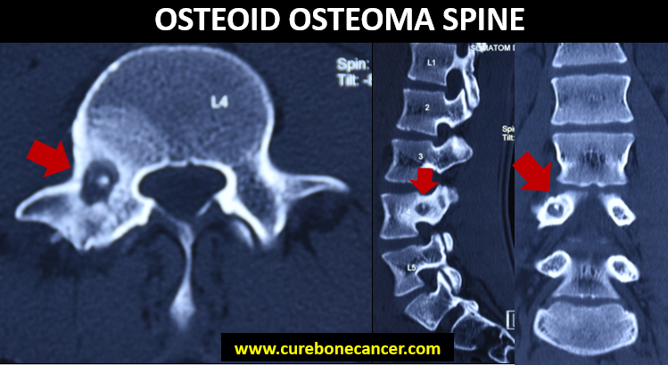
Osteoid osteoma in spine can cause spine deformity due to prolonged muscle spasm

Why has osteoid osteoma affected me?
There is no known cause for the development of osteoid osteoma. It is not linked to any dietary factors. Many patients attribute it to trauma, but that can merely be a coincidence.
What are the bones commonly affected by Osteoid osteoma?
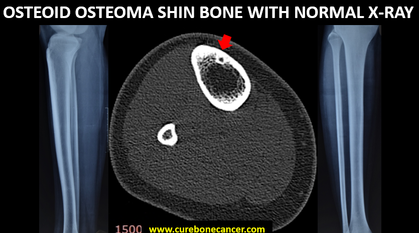
Osteoid Osteoma usually involves tibia (Shin Bone), Femur (Hip Bone) or spine, although it can involve any bone.

How is Osteoid Osteoma diagnosed?
A person suspected to have osteoid osteoma is subjected to series of investigations which include
- A plain X-ray is the first line modality for diagnosis of osteoid osteoma which may show eccentric periosteal thickening and sometimes a nidus, however it may be normal in many patients, thereby delaying the diagnosis and treatment. Typical history of night pain and relief with pain killers should prompt the treating clinician to get further investigations to rule out osteoid osteoma.
- A non contrast CT scan. This is the gold standard investigation for diagnosis of Osteoid Osteoma where the clinician identifies the pathognomic “nidus”.
- Dynamic contrast enhanced MRI. In doubtful cases, where a CT scan does not yield a diagnosis Dynamic Contrast enhanced MRI can be helpful. In this patient is given one injection of contrast and it’s pattern of uptake and washout from the lesion is measured. A rapid uptake and rapid washout clinches the diagnosis. Sometimes, a “nidus “ can also be localised on MRI only. Sometimes, because of diffuse edema around the lesion, it may not be easy to pick up the nidus only on plain MRI and hence, it is advised to get a specific Dynamic Contrast enhanced MRI at a specialised centre.
- Bone Scan. It is another modality to pick up osteoid osteoma in doubtful cases. A “ double density “ sign is pathognomic of osteoid osteoma on bone scan.
In majority of the cases, diagnosis of osteoid osteoma is confirmed just by a plain X-ray and a dedicated CT Scan.
Can osteoid osteoma mimic other diseases?
Osteoid osteoma may look like cortical osteomyelitis (infection) or stress fracture or rarely Ewing’s Sarcoma on radiological investigations and careful assessment by your treating Orthopaedic Oncologist can help differentiate one from the other
How is Osteoid Osteoma treated? Does it require a major surgery?
The main aim of treating osteoid osteoma is to remove the pain causing “nidus”.
Contrary to popular belief, Osteoid osteoma does not require any big surgery or major incisions for it’s treatment. Rather, since osteoid osteoma is a small lesion, open surgery may sometimes remove normal bone leaving behind the nidus or excessive removal of normal bone may make the patient prone to iatrogenic fractures and hence minimally invasive procedures to remove the nidus form the current standard of care.
Minimally invasive procedures (which require only one to two stitches) include :
- CT guided “Percutaneous Drilling” , in which nidus is localised under CT scan guidance and removed with the help of drill bits
- CT guided Radio Frequency Ablation (RFA) is another minimally invasive technique used for treating such lesions in which the lesion is burnt with thermal probe inserted into the nidus and burnt at 90 degree Celsius for 8 – 10 minutes.
- CT guided cryoablation is also done in a minimally invasive fashion in which the lesion is cooled to -70 degree celsius
- All these procedures can be done without hospital admission as a day care procedure.
Are procedures used to treat osteoid osteoma painful?
All these procedures , be it CT guided percutaneous drilling or Radiofrequency ablation, are done under general anaesthesia so that patient doesn’t experience any pain during the procedure. And in almost all the cases, the pain killer requirement reduces drastically to almost nil within a couple of days post the CT guided percutaneous drilling or Radiofrequency Ablation procedure
Is there any role of non surgical treatment in osteoid osteoma?
Osteoid osteomas can resolve on their own but it may take several years and patient has to take pain killers all this while. Taking over the counter pain killers for long have their own side effects and hence, non surgical treatment for osteoid osteoma is not recommended. Moreover, with the current techniques and expertise the minimally invasive procedures like CT Guided percutaneous drilling or CT Guided Radio Frequency Ablation has no downtime and can easily be done as a day care procedure without requiring any prolonged hospital stay.
What is follow up protocol after procedure for osteoid osteoma is done?
Patient needs to visit the doctor couple of times post procedure over next three months. Usually it’s done to assess healing of the tissues and monitor the symptoms if any.
What are the chances of tumour coming back after the treatment is over?
The chance of osteoid osteoma coming back is less than 5%. However, if it recurs, it can again be treated with percutaneous drilling or radio frequency ablation
Whom to consult when diagnosed with osteoid osteoma ?
It’s advisable to consult a specialist in bone tumours (Orthopaedic Oncologist) for optimal results.
Dr Rajat Gupta
Orthopaedic Oncologist
Bone Cancer Clinic
# 5, Sector 19 A, Chandigarh
https://goo.gl/maps/KT5hkxsRVy12
SCO 141 (Backside), Sector 14, Panchkula
https://goo.gl/maps/aLdmMwMqvMumVXe76